 |
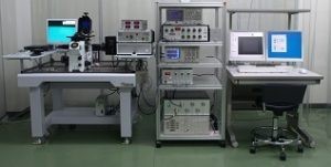
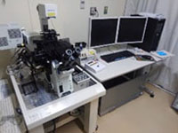
B001: ニコン 高速共焦点顕微鏡 A1R
- 生細胞共焦点蛍光イメージング
- レゾナントスキャナーとピエゾ Z ステージスキャナーによる高速共焦点観察
- 4波長レーザーライン (405 nm、488 nm、561 nm、640 nm)
- 32チャンネル分光検出ユニットによるスペクトル分離画像取得
- 導入年度: 2007
[ Detail ]
- ECLIPSE Ti-E (倒立型)
- PerfectFocus ユニット (長時間の生細胞観察で焦点面を保持)
- 電動 XY ステージ
- ピエゾ Z スキャナー
- 落射蛍光ユニット (Intensilight プリセンタードファイバー照明)
- 微分干渉コントラスト (DIC) 観察ユニット
- レーザー: 405 nm、488 nm、561 nm、640 nm
- 対物レンズ:CFI Plan Apo 10×、CFI Plan Apo VC 20×、CFI S Plan Fluor ELWD 20×C、CFI Plan Apo Lambda D 40×、CFI Plan Apo VC 60× (油浸)、CFI Plan Apo VC 60× (水浸)
- 落射蛍光フィルターセット: DAPI、FITC、TRITC
- 高速レゾナントスキャナー (7.8 kHz): 30 fps (512×512 px) および 420 fps (512×32 px) での高速撮影
- GaAsP 検出器 2 基 (488 nm・561 nm チャンネル用)、PMT 検出器 2 基 (405 nm・640 nm チャンネル用)、PMT 検出器 1 基 (微分干渉用)
- 32 チャンネル分光検出ユニット 1 基:1.67 fps (512×512 px) および 24 fps (512×32 px) でのスペクトル分離イメージング
- 蛍光フィルター:450/50、482/35、525/50、540/30、595/50、700/75
- 温度/CO₂ 制御付き細胞培養チャンバー (Tokai Hit 製)
|
 |
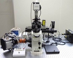
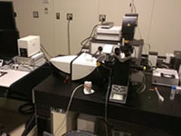
B002: オリンパス 共焦点超解像顕微鏡 FV-1000
- 生細胞共焦点蛍光イメージング
- FV-OSR 2D 超解像イメージング (xy 分解能 120 nm)
- 6 波長レーザーライン (405 nm、440 nm、488 nm、515 nm、559 nm、635 nm)
- 導入年度: 2007
[ Detail ]
- IX81ZDC オートフォーカスユニット付き電動倒立顕微鏡
- 電動 XY ステージ
- 落射蛍光ユニット (水銀ランプ照明)
- 微分干渉コントラスト (DIC) 観察ユニット
- レーザー: 405 nm、440 nm、488 nm、515 nm、559 nm、635 nm
- 対物レンズ: UPLSAPO 10×、UPLSAPO 20×、UPLSAPO 40×、UPLSAPO 60× (油浸)、UPLSAPO 100× (油浸)、UPLSAPO30XS (シリコン浸)
- 落射蛍光フィルターセット: DAPI、FITC、TRITC
- PMT 検出器 4 基、GaAsP 検出器 1 基
- 超解像ユニット/ソフトウェア FV-OSR
- 温度/CO₂ 制御付き細胞培養チャンバー (Tokai Hit 製)
|
 |
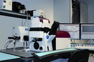
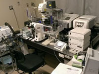
B003: ライカ STED超解像顕微鏡 TCS SP8 STED
- 生細胞共焦点蛍光イメージング
- ゲート付き STED 2D 超解像イメージング (xy 分解能 50 nm)
- 5 波長レーザーライン (458 nm、476 nm、488 nm、514 nm、633 nm) およびホワイトライトレーザー (470–670 nm)
- 2波長の STED レーザー (592 nm、660 nm) による 1〜3 色 STED イメージング
- 導入年度: 2009
[ Detail ]
- SP8 電動倒立共焦点顕微鏡 (Leica DMI6000 CS ベース)
- Adaptive Focus Control ユニット (長時間観察で焦点面を保持)
- レーザー: Ar (458 nm、476 nm、488 nm、514 nm)、HeNe (633 nm)
- ホワイトライトレーザー: 470–670 nm (AOTF により最大 8 波長同時選択可、80 MHz パルス)
- 連続波STEDレーザー (592 nm、660 nm) による STED
- Ti:Sapphire パルスレーザー (Coherent Chameleon)
- 検出器: PMT 蛍光検出器 2 基、ハイブリッド GaAsP 検出器 HyD 2 基 (時間ゲート検出により xy 分解能 <50 nm)、透過光検出器 1 基
- スキャナー: デュアルガルボミラー FOV スキャナー (高速 7 枚/秒 〈512×512 px〉、高密度 8k×8k 〈64 メガピクセル〉)
- 電動 XY ステージ/Z ガルボステージによる高精度 Z 走査
- 微分干渉コントラスト (DIC) 観察ユニット
- 対物レンズ: HC PL APO CS 10×/0.40 (ドライ)、HCX PL APO CS 40×/1.25–0.75 (油浸)、HCX PL APO CS 63×/1.40–0.60 (油浸)、HC PL APO 100×/1.40 STED WHITE (油浸)、HCX PL APO 63×/1.20 W CORR CS (水浸)
- 温度/CO₂ 制御付き細胞培養チャンバー (Tokai Hit 製)
- STED 対応デコンボリューションソフトウェア (Huygens)
|
 |
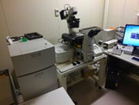
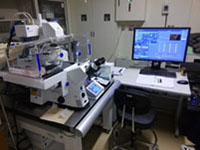
B004: ツァイス 共焦点・FCCS顕微鏡 LSM780
- 長時間生細胞共焦点蛍光イメージング
- 7 波長レーザーライン (405 nm、440 nm、458 nm、488 nm、514 nm、561 nm、633 nm)
- 32 チャンネル GaAsP 検出器アレイによるスペクトル分離画像取得
- FCS/FCCS (ConfoCor3+APD 検出器) による分子間相互作用解析
- 導入年度: 2009
[ Detail ]
- Axio Observer.Z1 電動倒立顕微鏡
- レーザー: 405 nm、440 nm、458 nm、488 nm、514 nm、561 nm、633 nm
- 検出器: QUASAR GaAsP 分光検出器 34 チャンネル、透過光検出器 1 基、多光子用外部検出器 2 基 (GFP 用・RFP 用)、FCS/FCCS 用 APD 検出器 2 基
- Definite Focus オートフォーカスユニット
- 電動 XY ステージ
- 落射蛍光ユニット (X-Cite ファイバー光源)
- 微分干渉コントラスト (DIC) 観察ユニット
- 対物レンズ: Plan-Apochromat 10×/0.45、Plan-Apochromat 20×/0.80、LCI Plan-Neofluor 25× Imm/0.80、C-Apochromat 40× Water/1.20、Plan-Apochromat 63× Oil/1.40
- 温度/CO₂ 制御付き細胞培養チャンバー (インキュベーションチャンバー+ヒーター+ CO₂ 供給)
- マイクロマニピュレーター (Narishige 製)
|
 |
B005: ツァイス 超解像顕微鏡 LSM880 Airyscan
- 長時間生細胞共焦点/超解像イメージング
- 6 波長レーザーライン (405 nm、458 nm、488 nm、514 nm、561 nm、633 nm)
- Airyscan 超解像イメージング (xy 分解能 120 nm、z 分解能 350 nm)
- Airyscan 高速モード (4 倍速撮影)
- 32 チャンネル GaAsP 検出器アレイによるスペクトル分離画像取得
- 導入年度: 2015
[ Detail ]
- Axio Observer.Z1 電動倒立顕微鏡
- レーザー: 405 nm、458 nm、488 nm、514 nm、561 nm、633 nm
- 検出器: QUASAR GaAsP 分光検出器 34 チャンネル、透過光検出器 1 基、高速撮影対応 Airyscan 検出器(4 倍速)
- Definite Focus オートフォーカスユニット
- 電動 XY ステージ
- ピエゾ Z ステージ
- 落射蛍光ユニット (HXP120V ファイバー光源)
- 微分干渉コントラスト (DIC) 観察ユニット
- 対物レンズ: Plan-Apochromat 10×/0.45、Plan-Apochromat 20×/0.80、LCI Plan-Neofluor 25× Imm/0.80、Plan-Apochromat 40× Oil/1.30、Plan-Apochromat 40× Oil/1.40、Plan-Apochromat 63× Oil/1.40 (超解像用)、Plan-Apochromat 100× Oil/1.46 (超解像用)
- 温度/CO₂ 制御付き細胞培養チャンバー (インキュベーションチャンバー+ヒーター+ CO₂ 供給)
|
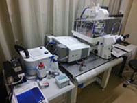 |
B006: ツァイス 共焦点顕微鏡 LSM710
- 長時間生細胞共焦点蛍光イメージング
- 5 波長レーザーライン (458 nm、488 nm、514 nm、561 nm、633 nm)
- 複数視野のタイリング/スティッチング
- 導入年度: 2009
[ Detail ]
- レーザー: 458 nm、488 nm、514 nm、561 nm、633 nm
- 検出器: QUASAR 分光検出器 2 チャンネル、LSM BiG GaAsP 検出器 2 チャンネル(GFP 用・RFP 用)、透過光検出器 1 基
- Definite Focus オートフォーカスユニット
- 電動 XY ステージ
- 落射蛍光ユニット (X-Cite ファイバー光源)
- 微分干渉コントラスト (DIC) 観察ユニット
- 対物レンズ: Plan-Apochromat 10×/0.45、LD LCI Plan-Apochromat 25× Imm/0.80、Plan-Apochromat 40× Oil/1.40、LD C-Apochromat 40× Water/1.10、Plan-Apochromat 100× Oil/1.46
- 温度/CO₂ 制御付き細胞培養チャンバー (インキュベーションチャンバー+ヒーター+ CO₂ 供給)
|
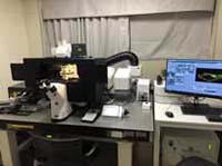 |
B007: ツァイス 多光子超解像高速顕微鏡 LSM980NLO Airyscan2
- 長時間生細胞共焦点/超解像/2 光子イメージング
- 7 波長レーザーライン (405 nm、445 nm、488 nm、514 nm、561 nm、639 nm、Chameleon Ultra II 680–1080 nm)
- Airyscan 2 超解像イメージング (xy 分解能 120 nm、z 分解能 350 nm)
- Airyscan 2 高速・低光毒性マルチプレックスモード (4 倍速・8 倍速撮影)
- 32 チャンネル GaAsP 検出器アレイによるスペクトル分離画像取得
- 導入年度: 2019
[ Detail ]
- Axio Observer.7 電動倒立顕微鏡
- レーザー: 405 nm、445 nm、488 nm、514 nm、561 nm、639 nm、Coherent Chameleon Ultra II (680–1080 nm)
- 検出器: QUASAR GaAsP 分光検出器 34 チャンネル、透過光検出器 1 基、マルチプレックス (4 倍速・8 倍速) 対応 Airyscan 2 検出器
- Definite Focus.2 オートフォーカスユニット
- 電動 XY ステージ
- ピエゾ Z ステージ
- 落射蛍光ユニット (HXP120V ファイバー光源)
- 微分干渉コントラスト (DIC) 観察ユニット
- 対物レンズ: Plan-Apochromat 10×/0.45、Plan-Apochromat 20×/0.80、Plan-Apochromat 63× Oil/1.40 (超解像用)、alpha Plan-Apochromat 63× Oil/1.46 (超解像用)
- 温度/CO₂ 制御付き細胞培養チャンバー (インキュベーションチャンバー+ヒーター+ CO₂ 供給)
- Arivis Vision 4D 画像処理ソフトウェア
|
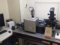 |
B008: アンドール 高速共焦点顕微鏡統合システム Dragonfly
- Borealis 均一照明によるスピニングディスク共焦点イメージング
- 最大 400 fps の高速生細胞イメージングと大型試料の 3D イメージング
- 4 波長レーザーライン (405 nm、488 nm、561 nm、637 nm)
- 高感度カメラ 2 基(iXon Ultra 888 EMCCD、Zyla 4.2 Plus sCMOS)
- Mosaic3 光刺激ユニット (405 nm レーザー)
- 導入年度: 2019
[ Detail ]
- Dragonfly 502: ピンホール径 25 µm/40 µm 切替式スピニングディスク、Borealis 照明、レーザー照明ワイドフィールドモード、カメラポート 2 基、3 段階電動カメラ倍率、イルミネーションズーム、画像取得と並行したGPU アクセラレーテッドデコンボリューション、Imaris Core
- Nikon Ti2-E 電動倒立顕微鏡
- レーザー: 405 nm/100 mW、488 nm/150 mW、561 nm/100 mW、637 nm/140 mW
- カメラ: iXon Ultra EMCCD、Zyla 4.2 Plus sCMOS
- Perfect Focus オートフォーカスユニット
- ASI 電動 XY ステージ&ステージピエゾ (可動範囲 500 µm)
- 対物レンズ:
- CFI Plan Apochromat Lambda 10× Dry NA 0.45 WD 4.00
- CFI Plan Fluor 20× M Imm (Oil/Glycerin/Water) NA 0.75 WD 0.35–0.33
- CFI Plan Apochromat Lambda S 25× Silicone NA 1.05 WD 0.55
- CFI Plan Apochromat Lambda S 40× Silicone NA 1.25 WD 0.30
- CFI Plan Apochromat VC 60× Water NA 1.20 WD 0.31–0.28
- CFI Apochromat TIRF 60× Oil NA 1.49 WD 0.13–0.07 (37 °C)
- CFI Apochromat TIRF 100× Oil NA 1.49 WD 0.15–0.09 (37 °C)
- CFI Plan Apo Lambda 60× Oil NA 1.40 WD 0.13
- CFI Plan Apo VC 100× Oil NA 1.40 WD 0.13
- Tokai Hit ステージトップインキュベーター
- Mosaic3 光刺激ユニット (405 nm/450 mW レーザー)
|
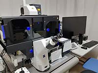 |
B009: ツァイス 格子構造化照明超解像顕微鏡 Elyra7 with Lattice SIM^2
- 最速 255 fps の生細胞超解像イメージング
- 4 波長レーザーライン (405 nm、488 nm、561 nm、642 nm)
- 高光効率の格子構造化照明 & 高度デコンボリューション (Lattice SIM^2) (xy 分解能 60 nm、z 分解能 200 nm)
- 高速スライス画像取得用 SIM^2 Apotome モード
- 高感度カメラ 2 基 (PCO edge 4.2 sCMOS) による同時 2 色撮影
- 導入年度: 2021
[ Detail ]
- Axio Observer.7 電動倒立顕微鏡
- レーザー: 405 nm/50 mW、488 nm/100 mW、561 nm/100 mW、642 nm/150 mW
- カメラ: PCO edge 4.2 sCMOS ×2 (Duolink モジュールに設置)
- Definite Focus.2 オートフォーカスユニット
- 電動 Z ドライブ
- ピエゾ走査ステージ: XY 130 × 100 mm、Z 100 µm
- 落射蛍光ユニット (HXP120V ファイバー光源)
- 微分干渉コントラスト (DIC) 観察ユニット
- 対物レンズ: EC Plan-Neofluar 10×/0.30、LD LCI Plan-Apochromat 25× Imm Corr/0.80、Plan-Apochromat 40× Oil/1.40、alpha Plan-Apochromat 100× Oil/1.46
- 温度/CO₂ 制御付き細胞培養チャンバー (インキュベーションチャンバー+ヒーター+ CO₂ 供給)
|
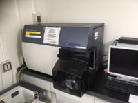 |
B101: BD フローサイトメーター Canto II
- 2本のレーザーを搭載 (L1: 青 488 nm, L2: 赤 633 nm)
- 6色を同時測定可能(対応蛍光色素やフィルター等構成の詳細は下記参照。)
- FACSDiva 8 制御ソフトフェアを使用
- 98穴または384穴プレートを利用可能(ハイスループットサンプラー(HTS)付)
- 導入年度: 2008
[ Detail ]
- 青 A: PE-Cy7 (735 LP, 780/60)
- 青 B: PerCP-Cy5-5 (655LP, 670LP)
- 青 C: (610LP)
- 青 D: PE (556LP, 585/42)
- 青 E: FITC (502LP, 530/30)
- 青 F: SSC (488/10)
- 赤 A: APC-Cy7 (735LP, 780/60)
- 赤 B: (685LP)
- 赤 C: APC (660/20)
|
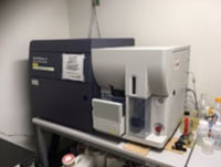 |
B102: BD セルソーター Aria II SORP
- 4本のレーザーを搭載 (L1: 青 488 nm, L2: 赤 640 nm, L3: 紫外 355 nm, L4: 緑 532 nm)
- 13色同時測定可能(対応蛍光色素やフィルター等構成の詳細は下記参照。)
- FACSDiva 8 制御ソフトウェアを使用
- プレートソーティング対応
- サンプル冷却システム付
- 導入年度: 2009
[ Detail ]
- 青 A: PE-Cy7 (750 LP, 780/60)
- 青 B: PerCP-Cy5-5 (685LP, 710/50)
- 青 C: PE-Cy5 (635LP, 660/20)
- 青 D: PE-Texas Red (600LP, 610/20)
- 青 E: PE (550LP, 575/25)
- 青 F: FITC (505LP, 525/50)
- 赤 A: APC-Cy7 (750LP, 780/60)
- 赤 B: Alexa Fluor 700 (690LP, 730/45)
- 赤 C: APC (660/20)
- 紫外 A: Hoechst Red (635LP, 670/50)
- 紫外 B: Hoechst Blue (450/40)
- 緑 A: Grn PE-Cy7 (750LP, 780/60)
- 緑 B: Grn PE (575/25)
|
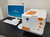 |
B104: ProteinSimple キャピラリーイムノブロッティング Jess System
- キャピラリー化学発光および蛍光ウェスタンブロッティング装置
- 2-40 kDaまたは12-230 KDa、 66-440 kDaの分子量分離が可能な13本または25本のキャピラリーを有するカートリッジを利用可能
- 導入年度: 2020
[ Detail ]
|
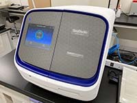 |
B105: Applied Biosystems 4キャピラリーDNAシーケンサー Seqstudio Genetic Analyzer
- サンガー法
- 4本のキャピラリーを搭載
- サーモフィッシャーコネクト対応(自宅からの遠隔状況確認が可能)
- 導入年度: 2020
[ Detail ]
- 単一プラットフォームでサンガー法とフラグメント解析が可能な4キャピラリー電気泳動システム。
- ポリマー、バッファー、キャピラリーアレイを一体化した試薬カートリッジベースのシステム。
- 柔軟なサンプルスループット、自動サンプル注入により1~4サンプル/ランをサポート。
- 直感的なワークフローを実現する最新のタッチスクリーンインターフェース。
- 付属のデータ解析ソフトウェア:SeqStudio Analysis Software、GeneMapper、Varian Reporter など
|
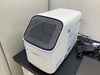 |
B106: Applied Biosystems RT-PCR装置 QuantStudio 3
- 精密な温度制御と最適化が可能な3つの独立した温度帯を搭載
- 標準的な96ウェルプレートに対応(MicroAmp Optical 96ウェル反応プレート(カタログ番号:N8010560)またはMicroAmp Optical 8連チューブストリップ(0.2 mL、カタログ番号:4316567)に対応)
- サーモフィッシャーコネクト対応(自宅からの遠隔状況確認が可能)
- 導入年度: 2021
[ Detail ]
- 高速および標準サイクリングオプションを備えた96ウェルブロックフォーマットにより、ハイスループットで柔軟な実験ワークフローを実現。
- 4つの光学チャネルによるマルチプレックス機能により、1回の反応で複数のターゲットを同時に検出。
- 直感的なタッチスクリーンインターフェースとガイド付きワークフローにより、ユーザーフレンドリーな操作性と最小限のトレーニングで利用可能。
- Thermo Fisher Connectとのクラウド対応接続により、リモートでの実験セットアップ、モニタリング、データ解析が可能。
- 高い感度と再現性を備え、遺伝子発現、ジェノタイピング、病原体検出などのアプリケーションに最適。
|
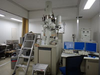 |
M001: 日本電子 電界放出形透過電子顕微鏡(FE-TEM) JEM-2200FS+JED-2300T
- ナノスケールにおける形態観察や構造解析などの局所状態分析
- EDXによるナノ領域の元素分析
- オメガ型エネルギーフィルターを用いた高コントラスト観察
- 低温・高温試料ホルダー
- 導入年度: 2009
[ Detail ]
- 分解能:格子分解能0.05nm、点分解能0.14nm(200kV)
- TEM(BF、DF)、SAED、STEM(BF、HAADF)、EDX(点、線、マッピング)、EELS
|
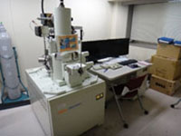 |
M002: 日本電子 電界放出形走査電子顕微鏡(FE-SEM)JSM-7500F
- ナノ - マイクロスケールの形状観察および組成分析
- 導入年度: 2008
[ Detail ]
- 分解能:1.0nm(15kV)、1.4nm(1kV)
- 加速電圧:0.1kV~30kV
- 倍率:25倍~1,000,000倍
- 方式:低加速電圧モード(ジェントルビーム)、エネルギーフィルター(ガンマフィルター)
|
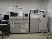 |
M003: リガク X線吸収分析装置 XAS-Looper
- 試料中の元素組成および構造の分析
- 導入年度: 2008
[ Detail ]
- 最大3kW
- 非破壊分析
- 元素選択性
- 特定元素における局所構造分析
|
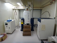 |
M004: ブルカー 核磁気共鳴装置(NMR)AVANCE-III 500USP
- 溶液中の有機分子の分子構造解析
- 導入年度: 2009
[ Detail ]
- 共鳴周波数(1H) 500 MHz
- プローブ 1H/19F & 31P~15N 5mm オートチューニングモード
- オートサンプラー(最大24サンプル)
- 温度可変測定 -20℃~+100℃
|
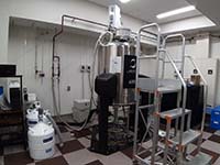 |
M006: 日本電子 核磁気共鳴装置 (NMR) JNM-ECZ600R/M3
- 600 MHz固体NMRシステム(24サンプルASC搭載)
- 導入年度: 2019
[ Detail ]
- CZR型固体NMR分光計(中出力アンプ搭載)
- <プローブ>1 mm HX MAS/広温度範囲
- 3.2 mm HX MAS/8 mm HX MAS/3.2 mm AutoMAS/
- GR(高勾配磁場、固体用サンプルチューブ)/5 mmFG/ROYALHFX
|
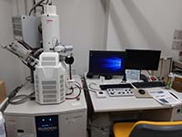 |
M007: 日立ハイテクノロジーズ
ショットキー走査電子顕微鏡 SU5000
- ナノからマイクロスケールの形状観察と組成分析
- 導入年度: 2014
[ Detail ]
- 分解能 1.2 nm(30kV)、3.0 nm(1kV)
- 加速電圧 0.5 kV~30 kV
- 倍率 25~60万倍
- エネルギー分散型X線分光装置(EDS)、大面積観察
|
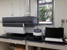 |
M008: 島津製作所 ICP発光分析装置 ICPE-9820
- 水溶液に溶解した試料中の元素分析
- 高感度:微量元素も検出可能 (ppm~ppb:元素により異なる)
- 多元素分析
- 導入年度: 2024
[ Detail ]
- 水溶液に溶解した試料中の元素分析
- 高感度:微量元素も検出可能 (ppm~ppb:元素により異なる)
- 多元素分析
|
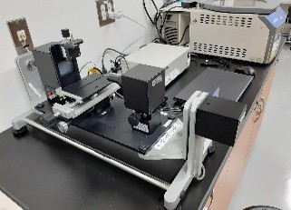 |
M009: 協和界面科学 自動接触角計 DMs-401
- 静的および動的接触角、固体の表面自由エネルギー、液体の表面張力
- および界面張力を測定するための小型高性能表面測定装置。
- 導入年度: 2015
[ Detail ]
- 動的濡れ挙動を高速・高精度に観察:DMs-401は、60fpsの高速CMOSカメラを搭載し、液滴挙動をリアルタイムで可視化します。これにより、初期の広がり、吸収、界面活性剤効果といった時間依存現象の詳細な解析が可能になります。
- 包括的な評価を可能にする多彩な測定機能:本システムは、静的および動的接触角測定(前進/後退)、表面張力および界面張力測定(ペンダントドロップ法)、表面自由エネルギー解析(OWRK法、Zisman法など)をサポートしています。幅広い用途における濡れ性、接着性、表面処理の評価に最適です。
- 拡張性を実現するモジュール設計:DMs-401は、温度制御ユニット、自動ディスペンサー、外部傾斜ステージなどのオプションを追加することで、進化する測定ニーズに合わせてカスタマイズできます。柔軟なアーキテクチャにより、将来を見据えた使いやすさを実現します。
|
 |
M010: リガク 電子線回折構造解析装置
- TEM(JEOL)とSCXRD(Rigaku)を1台に統合
- 粒子サイズ範囲:数十nm~最大1,000nm(1µm)、XRDで使用される単結晶の約100倍小さい
- 画像取得モードのシームレスな切り替え
- 高速測定:1セットの回折データ取得には数分~30分かかります。
- 導入年度: 2024
[ Detail ]
- ナノサイズ結晶の構造解析が可能:XtaLAB Synergy-EDは、数十ナノメートルから数百ナノメートルという極めて小さな結晶の構造解析を可能にします。従来のX線回折では解析できなかった試料の解析に新たな可能性をもたらします。
- 高速でシームレスなワークフロー:試料の選択から回折データの収集、構造解析まで、「CrysAlisPro for ED」によってプロセス全体が合理化されます。これにより、最小限の手作業で効率的かつ迅速な構造解析が可能になります。
- 直感的な操作 - 専門知識は不要:電子顕微鏡の使用経験がない研究者でも、単結晶X線分析の知識があれば、XtaLAB Synergy-EDを初日から使用できます。3D電子回折(3DED / MicroED)技術をすぐに活用できます。
|
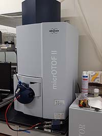 |
M011: ブルカー 大気圧化学イオン化/エレクトロ
スプレーイオン化飛行時間型質量分析計
(APCI/ESI-TOF MS) micrOTOF II
- 飛行時間型質量分析計(TOF MS)システム
- 選択可能なイオン化法:大気圧化学イオン化(APCI)またはエレクトロスプレーイオン化(ESI)
- 用途:メタボロミクス、創薬、ポリマー分析
- 導入年度: 2013
[ Detail ]
- 15,000 FWHMを超える分解能を備えた高分解能質量分析計により、複雑な混合物の精密質量測定が可能。
- 内部キャリブレーションにより2ppm未満の質量精度を実現し、信頼性の高い分子式決定が可能。
- UHPLCシステムとの連携に適した高速データ取得速度により、高速クロマトグラフィーピークのリアルタイム分析が可能。
- m/z 3000までの広い質量範囲により、低分子、ペプチド、その他の生体分子の検出可能。
|
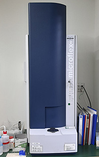 |
M012: ブルカー マトリックス支援レーザー脱離イ
オン化飛行時間型質量分析計(MALDI-TOF
MS) Microflex Reflectron-KS II
- 飛行時間型質量分析計(TOF MS)システム
- イオン化法:マトリックス支援レーザー脱離イオン化法
- 用途:タンパク質およびペプチド分析、バイオマーカー探索、ポリマーおよび合成材料分析
- 導入年度: 2009
[ Detail ]
- Reflex™イオン光学系は、遅延抽出法とリフレクトロンTOF技術により、高い質量分解能と精度を実現。
- リニアモードとリフレクトロンモードをサポートし、タンパク質などの高分子からペプチド、代謝物などの低分子化合物まで、柔軟な分析が可能。
- 高ショット周波数の堅牢な窒素レーザー(337 nm)により、迅速なデータ取得と高い再現性を実現。
- コンパクトなベンチトップ設計で、省スペースで高性能なMALDI-TOF MSソリューションを必要とする研究室に最適。
|
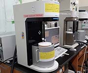 |
M103: 吸着装置 BELSORP-MINI X
- ガス吸着・脱着 (N2, CO2, O2, Ar)
- 導入年度: 2021
[ Detail ]
- 77 K (液体窒素), 0~40 ˚C
- BET 表面積
|
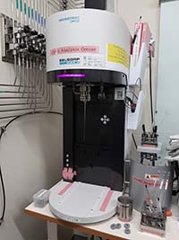 |
M104A: 吸着装置 BELSORP-MAX X-A
- 幅広い材料を高精度に測定
- 比表面積0.01 m²/g (N₂/Ar)から0.0005 m²/g (Kr)まで、細孔径分布0.35 nmから500 nmまでをカバー。
- 活性炭、触媒、セラミックスなどの特性評価。
- 測定時間短縮:従来法と比較して測定時間を約50~70%短縮
- 高度な分析ソフトウェア:BET 表面積、細孔分布、吸着速度論分析など
- 導入年度: 2010, 2012
[ Detail ]
- ガス吸着・脱着(He、N₂、O₂、CO₂、Ar)
- H₂O、有機溶媒対応可能
- 77K(液体窒素)、0~40℃
- BET比表面積
- 極低圧下での測定
- 細孔分布分析
|
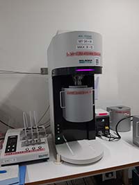 |
M104B: 吸着装置 BELSORP-MAX X-B
- 幅広い材料を高精度に測定
- 比表面積0.01 m²/g (N₂/Ar)から0.0005 m²/g (Kr)まで、細孔径分布0.35 nmから500 nmまでをカバー。
- 活性炭、触媒、セラミックスなどの特性評価。
- 測定時間短縮:従来法と比較して測定時間を約50~70%短縮
- 高度な分析ソフトウェア:BET 表面積、細孔分布、吸着速度論分析など
- 導入年度: 2024
[ Detail ]
- ガス吸着・脱着(He、N₂、O₂、CO₂、Ar)
- H₂O、有機溶媒対応可能
- 77K(液体窒素)、0~40℃
- BET比表面積
- 極低圧下での測定
- 細孔分布分析
|
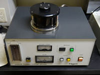 |
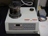
M201: フィルジェン オスミウムプラズマコーター OPC60AL
- SEM試料表面に導電性オスミウム薄膜を形成し、帯電を防止します。
- 粒度分布と熱ダメージを軽減します。
- 導入年度: 2009
|
 |
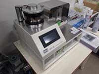
M202: 日本電子 オートファインコーター JFC-1600
- SEM試料表面に導電性Pt薄膜を形成し、帯電や熱による損傷を防止
- 導入年度: 2009
|
 |
M203: 真空デバイス
オスミウムプラズマコーター HPC-20
- SEM試料の表面に導電性オスミウム薄膜を形成し、帯電を防止します。
- 粒度分布と熱ダメージを低減します。
- 導入年度: 2014
|


